I. Laws of Pulp Chamber Location:
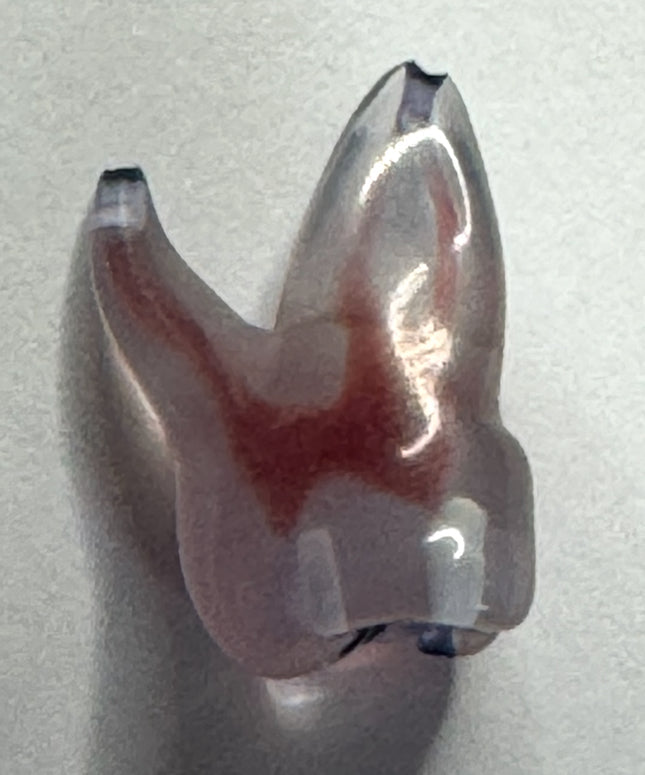
The Law of Centrality: The floor of the pulp chamber is consistently located in the geographic centre of the tooth at the level of the Cemento-Enamel Junction (CEJ).
The Law of Concentricity: The walls of the pulp chamber are concentric to the external surface of the tooth at the CEJ level. Consequently, the internal anatomy reflects the external root trunk morphology; if the CEJ bulges or narrows in a specific direction, the pulp chamber follows a corresponding path.
The Law of the CEJ: The CEJ serves as the most reliable anatomical landmark for determining the depth and position of the pulp chamber, regardless of coronal wear or restorative alterations.
II. Laws of the Pulp-Chamber Floor
The Law of Colour Change: The floor of the pulp chamber is significantly darker than the surrounding dentinal walls. This distinct visual contrast is a critical indicator for identifying the junction where the walls meet the floor.
The Law of Symmetry 1: In multi-canalled teeth (excluding maxillary molars), the orifices are equidistant from a line drawn in a mesio-distal direction through the centre of the pulp-chamber floor.
The Law of Symmetry 2: Orifices lie on a line perpendicular to the aforementioned mesio-distal midline. If a canal is located off-centre, a symmetrical counterpart is statistically likely to exist on the opposite side.
III. Laws of Orifice Location
Law of Orifice Location 1: Root canal orifices are invariably positioned at the junction of the pulp chamber walls and the floor.
Law of Orifice Location 2: Orifices are located at the specific angles formed by the floor-wall junction.
Law of Orifice Location 3: Orifices are positioned at the terminus of developmental root fusion lines (darker grooves on the chamber floor).
Adherence to these anatomical principles facilitates the transition from exploratory "blind" drilling to a disciplined, predictable clinical procedure.
- High Contrast: The vibrant blue dye provides a sharp visual contrast against the dentine of the pulp chamber floor.
- Diagnostic Precision: It is particularly effective for identifying elusive canals (like the MB2) and detecting developmental cracks that are invisible to the naked eye.
- Low Viscosity: The gel has a flowable texture that allows it to penetrate anatomical irregularities and secondary caries around old restorations.
- Controlled Application: Supplied in a 3ml syringe with 5 brush tips for targeted delivery with minimal waste
- Preparation: Remove any dentine or debris covering the expected orifice location.
- Application: Apply 1–3 drops of Canal-Seek into the bottom of the pulp cavity.
- Wait: Allow the dye to sit for 5–10 seconds to penetrate the orifices or cracks.
- Rinse & Dry: Thoroughly rinse the cavity with water and dry it. The areas stained intense blue indicate the canal openings.
- Clean Up: After use, pull back the syringe plunger to prevent overflow and recap the syringe securely
- Micro-Blade Technology: Unlike traditional diamond-coated tips, these use advanced micro-blades for precise cutting with reduced wear and improved durability.
- Built-in Irrigation: Features integrated water ports to keep the operative field clear and prevent overheating.
- High Visibility: The tips are engineered with specific angles (such as 110°) to allow for an incomparable view of the pulp chamber floor under magnification.
- Material: Made from high-strength surgical steel that is fully autoclavable.
The tips included are:
- AC1 (ED51): Used for finishing access cavity walls in molars and premolars to create a properly tapered shape.
- AC2 (ED52): Specifically designed for locating the MB2 canal and other difficult-to-find canal orifices.
- AC3 (ED53): Used for the removal of calcifications and fiber posts.
- AC4 (ED54): Designed for decrementing and removing metal posts.
- AC5 (ED55): Used for refining and cleaning the pulp chamber floor to expose canal anatomy
What is the recommended power?
Recommended Power: It is generally recommended to use these tips at 60% – 90% of the ultrasonic unit's power output.
at Toothsaver.co.uk are tungsten carbide round burs designed with extra-long, slim necks to provide an unobstructed view for identifying root canal orifices and preparing isthmuses. They are available as comprehensive kits or individual replacement burs in various sizes and lengths.
A full set containing 10 burs and a bur stand. The kit includes an assortment of sizes ranging from 004 to 014 in both 31mm and 34mm lengths.
Individual replacement burs:
- Size 004 (Purple): For fine exposure of the pulp chamber floor.
- Size 006 (White): Standard fine tracing.
- Size 008 (Yellow): For general tracing and isthmus preparation.
- Size 010 (Red): Removing excess dentine at canal entries.
- Size 012 (Blue): Recommended for long clinical crowns.
- Size 014 (Green): Larger diameter for initial orifice widening.
- Material: High-quality tungsten carbide with a particularly sharp cutting part for precise dentine removal.
This device functions as both a high-precision apex locator and an orifice finder. Unlike standard models, the MHN mode measures capacitance values to enhance signal detection sensitivity, making it easier to identify pulp chambers or orifices even when they are partially calcified.
Key Features:
- Dual Mode: Switch between standard Apex Locator (AL) mode and MHN mode.
- MHN Explorer Probe: Includes a specialised probe that can be connected to the device to "feel" for orifices electrically.
- Enhanced Processing: Features a digital circuit that processes information at double the speed of previous generations like the Woodpex V.
- Display: 3.8-inch LCD screen with an adjustable angle (up to 51.8 degrees) for better ergonomics.
Technical features:
- Battery: 3.7V / 2000mAh rechargeable lithium battery providing approximately 7 hours of continuous use.
- Charging: Requires 3 hours for a full charge via a standard adapter.
- Accuracy: Maintains high accuracy (reported at 97.71% or higher) even in canals containing blood or residual pulp.
Kit contents:
- 1 x Main Unit
- 1 x Measuring wire (non-autoclavable)
- 4 x File clips (autoclavable)
- 5 x Lip hooks (medical-grade stainless steel)
- 1 x MHN explorer probe (specifically for orifice location)
The Stropko™ Irrigator is a precision adapter designed to replace standard air/water syringe tips, allowing the use of small-gauge Luer-lock needles for controlled irrigation and drying.
The large variety of Luer-Lock tips enables direct access to any area of the mouth, during any procedure, no matter if it is the lingual of an anterior, the distal of a molar, or an apical retro-prep during surgery.
Using the Stropko Irrigator, cleaning and drying can be done with more precision and control, eliminating all unintentional splashing or contamination of the working area. With standard syringe tips, it is not possible to prevent the dentinal dust from obstructing the vision. A good example of vision control can be observed while troughing in search of hidden canals.