

Payment & Security
Your payment information is processed securely. We do not store credit card details nor have access to your credit card information.
Description
Apply today to join the workshop: Daniel@endouk.com as there are limited spaces. Payments by Bacs to account below (£1250) or contact us.
Endodontic Solutions Ltd
A/C 62148215
Sort: 608371
Iban: GB32SRLG60837162148215
Swift/bic: SRLGGB2L
We are proud to have Dr Mo Fayad and Dr Bruno attend “The Gathering” to deliver an intense 2 day world class workshop. You will need to bring your computer for day 2 during which you will use all your newly acquired knowledge to plan cases.
The 2 day workshop will be delivered by 2 experts in the field Dr Bruno Azevedo who will showcase how to utilise the power of enhanced software and on how to acquire and diagnose challenging pathology.
Dr Fayad will share his extensive experience in practice and how he incorporates the latest technology in his practice. He will share the clinical outcomes of how challenging pathologies have been treated with follow-ups and how his practice has changed over time.
This will be the first time this popular workshop has been hosted in Europe.
Several technologies have been introduced to the endodontic specialty in the past few years. Among these technologies are CBCT, dental lasers, AI softwares and dynamic navigation. CBCT has become the scaffold for other technologies. Importing CBCT dicom files into e-VOLDx artificial intelligence (AI) software and dynamic navigation systems have added a new dimension to diagnosis and treatment. The incorporation of CBCT technology and a new imaging software e-VOLDX has provided clinicians with 300 times higher resolution and the ability to visualize dental structures clinicians were not able to visualize in the past. Structures as the dental pulp, nerve bundles and maxillary sinus membranes are few of these examples. Dynamic navigation software allows the clinician to visualize dental procedure on a monitor in real time while performing challenging non-surgical and surgical procedures. Another technology are dental lasers which are utilized in non-surgical (acoustic streaming disinfection of root canal systems) and surgical procedures (laser guided surgical incision, soft tissue management in invasive cervical resorption and photo-biomodulation to accelerate wound healing).
This presentation will focus on how the incorporation of these technologies can be utilized in non-surgical and surgical challenging endodontic procedures. Based on the information provided, changes in treatment protocols and armamentarium will be presented.
Learning Objectives:
- Import CBCT dicom images into e-VOLDX AI software in the following cases: Diagnosis of pain, cracked teeth, vertical root fracture and resorptive defects.
- Apply dental lasers in non-surgical (acoustic streaming disinfection of root canal systems) and surgical procedures (laser guided surgical incision, soft tissue management in invasive cervical resorption and photo-biomodulation to accelerate wound healing).
A lack of consensus exists among dentists and endodontists on cracked teeth. Accurate clinical evaluation, imaging and diagnosis is paramount to properly treatment planning cracked teeth. With the resurgence of the philosophy of saving compromised teeth over extraction, and our patients’ desire to save their teeth, the question remains: “Which cracked teeth should be treated and which cracked teeth should be extracted?” Applying the current understanding of the ultrastructure of teeth in conjunction with modern diagnostic technologies, materials, adhesive techniques, and restorative approaches, dentists can now use minimally invasive biomimetic techniques to provide restorations that come close to functionally and biomechanically mimicking the original, healthy state of a tooth. This evidence-based presentation will address the classification and diagnosis of different types of cracks and the role of CBCT imaging in these cases, as well as the prognosis and changes in treatment protocols and outcomes of cracked teeth.
At the conclusion, participants should be able to:
- Describe the role of CBCT imaging in identifying cracked tooth types, extent of the fractures, and early bone pattern changes associated with cracked teeth.
- Explain the prognosis and outcomes studies related to treatment and management of cracked teeth and vertical root fractures.
- Apply the current understanding of the ultrastructure of teeth in conjunction with modern diagnostic technologies, materials, adhesive techniques, and restorative approaches for restoration of cracked teeth.
Real-time dynamic navigation and dental lasers are often hailed as groundbreaking innovations in various fields. Real-time dynamic navigation systems, particularly in non-surgical and surgical procedures provide enhanced precision and safety by continuously adapting to changing conditions. Dental lasers offer precise, minimally invasive treatments that can enhance patient outcomes and reduce recovery times. But are these technologies truly as transformative as they seem, or are they just advanced iterations of existing tools? This evidence based presentation will delve into the real impact of these innovations, assessing their effectiveness, adoption, and the extent to which they are changing the future non-surgical and microsurgical Endodontics. In this lecture, for the first time, the integration of real-time laser guidance with microsurgical techniques will be presented. Real-time laser guidance represents a significant leap in enhancing precision, safety, and outcomes in endodontic microsurgery.
Video footage of clinical cases will be utilized to demonstrate the 3-D scan evaluation as well as the surgical procedures for cases involving vital structures and cases where bone grafting is indicated.
At the conclusion, participants should be able to:
- Identify the importance of CBCT information in presurgical assessment, case selection and treatment planning.
- Utilize the appropriate Guided Bone Regeneration products as bone grafts and membranes in endodontic/periodontal and marginal defect cases when indicated.
- Utilize the CBCT information to identify and manage cases involving vital structure (maxillary sinus, inferior alveolar, mental and nasopalatine bundle).
Dr. Bruno Azevedo is a dual specialist in Endodontics and Oral Maxillofacial Radiology, currently in private practice in Austin, Texas. He has over 21 years of experience utilizing CBCT imaging, with a particular emphasis on Endodontics, and is the author of numerous papers and book chapters on this topic. He graduated from dental school at Gama Filho University- Rio De Janeiro, Brazil. He holds a Master's in Dental Diagnostic Sciences, a certificate in Oral Maxillofacial Radiology from the University of Texas Health Science Center in San Antonio, and a certificate in Endodontics from the Einstein Medical Center in Philadelphia. A Diplomate of the American Board of Oral and Maxillofacial Radiology, Dr. Azevedo is recognized as a leading North American expert in 3D imaging technologies for Endodontics. He was a founding faculty member of Western University College of Dental Medicine and the former director and division head at the University of Louisville. His passion for advancing the field through education is evident in his frequent speaking engagements worldwide.
Cone beam computed tomography (CBCT) is the newest, widely-adopted imaging modality available to the practicing endodontist. This discussion will review what available evidence says about the role of CBCT in endodontic case diagnosis, treatment planning, treatment outcomes, and practitioner confidence. We will also discuss the thought process and rational for ordering a CBCT scan and place into context the relevant risks of using ionizing radiation in the endodontics clinic.
At the conclusion, participants should be able to:
- Review what available evidence says about the role of CBCT in endodontics
- Explain the thought process and rationale for when to order at CBCT scan
- Place into context the relevant risks associated with use of ionizing radiation in an endodontics clinic
NS-3 - Overview of Medicolegal Aspects of Dental Radiology
Cone beam computed tomography (CBCT) has afforded the practicing endodontist a new realm of 3D diagnostic imaging with many clinical advantages, however; standing medicolegal principles of ordering any diagnostic test still apply. This discussion will review general principles of medicolegal aspects of dentistry including a presentation of the general thought process and rationale for ordering any radiographic examination in dentistry, a review of the general medicolegal duties of members of the dental team with respect to the standard of care in radiology, and discussion of image/scan reviewing and reporting responsibilities.
At the conclusion, participants should be able to:
- Describe the general thought process and rationale for ordering any radiographic examination in dentistry, including CBCT
- Review some general medicolegal duties of members of the dental team with respect to standard of care in radiology
- Discuss reviewing and reporting responsibilities
This presentation will cover the current and future applications of various advanced Imaging modalities in the endodontic practice. A live demonstration of future imaging integration using virtual environments such as the Meta-Verse - Virtual Reality (VR) and Augmented Reality (AR) will be demonstrate the profound impact of how newly developed technologies can help enhance diagnosis and increase collaboration between clinicians.
Learning Objectives:
- Update participants on current and future endodontic imaging technologies
- Explain how to use 3D Task-Specific Filters to detect anatomic variations and hard-to-see pathologies
- Describe how the Meta-Verse - Virtual Reality (VR) and Augmented Reality (AR) can help endodontists better diagnose complex clinical scenarios
Cone Beam Computed Tomography (CBCT) has become an indispensable diagnostic imaging tool in clinical endodontics. In particular, high resolution limited field of view 3D imaging volumes of the jaws provide endodontists with greater diagnostic accuracy for tooth and root morphology and both tooth and bone related pathology when compared to regular intraoral digital 2D imaging. Because the use of CBCT imaging provides clinicians with a greater degree of confidence in detection of anatomic variations and pathology, it acts as an important clinical decision support tool in that it provides valuable information on when to treat, how to treat and, just as importantly when not to treat. CBCT imaging is emerging as the scaffold for upcoming technologies such as 3D printing, dynamic navigation and surgical guides and is being applied to better understand the healing process associated with previously endodontically treated teeth. The American Association of Endodontists (AAE), in conjunction with the American Association of Oral and Maxillofacial Radiology (AAOMR), have developed and recently updated a position statement on the use CBCT imaging in endodontics providing evidence based clinical practice situations in which 3D imaging can be considered as the preferred imaging modality before, during and after endodontic therapy. Currently the position statement does not address best practices involving technical issues such as acquisition parameters and image display settings for optimizing image quality and maximizing the diagnostic capabilities of available CBCT units. Using clinical cases, we will demonstrate the effect of task specific enhancement filters and discuss in which clinical scenarios should they be applied and the impact of newly developed realistic 3D rendering for endodontic diagnosis.
At the conclusion of this presentation, participants will be able to:
- Be familiar with current technical advances in CBCT hardware and software;
- Understand Contrast Artifacts and how to overcome them
- Integrate information presented in this course into efforts to improve the diagnostic imaging skills of participants
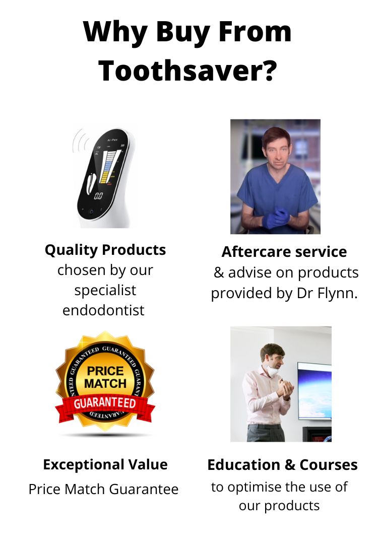
Ask Our Expert - Dr Daniel Flynn, Specialist Endodontist